Rotator Cuff Tear Mri With Contrast
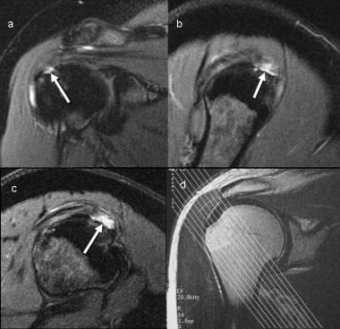
Indirect signs on mri are subdeltoid bursal effusion medial dislocation of biceps fluid along biceps tendon and diffuse loss of peribursal fat planes. On the right the same muscle is torn dark signal.
 Parial Rotator Cuff Tear Note Stripes Of Contrast Medium Near The
Ct arthrogram for patients who cannot have an mri proximal biceps injury mri shoulder without contrast.
Rotator cuff tear mri with contrast. A routine shoulder mri which takes about 25 minutes of actual scan time and shoulder mr arthrogram which involves an x ray procedure during which contrast is injected directly into the. The needle for the injection is visible in the shoulder in this ct image. Newer treatment options for rotator cuff tears include using your body s own repair cells to treat the damaged muscle or tendon using stem cell therapy.
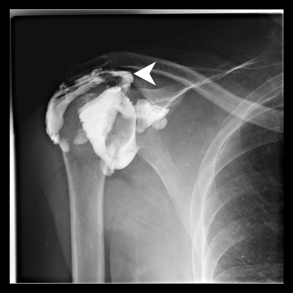
Us for patients who cannot have an mri. Tendon retraction may also be present which can be graded using the patte classification. A ct arthrogram should be specified in order to rule out a rotator cuff tear.
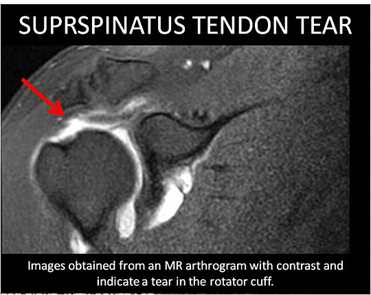
When only a partial thickness tear is present the scan will show an extension of the contrast agent into the substance of the tendon. The presence of a tendon defect filled with fluid is the most direct sign of rotator cuff tear. Although a rotator cuff tear won t show up on an x ray this test can visualize bone spurs or other potential causes for your pain such as arthritis.
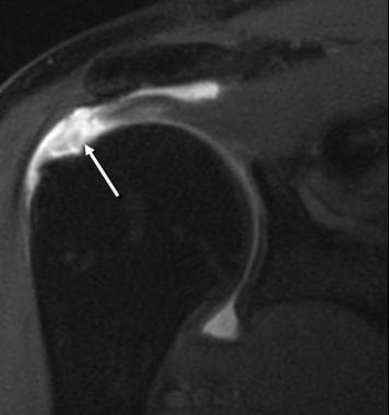
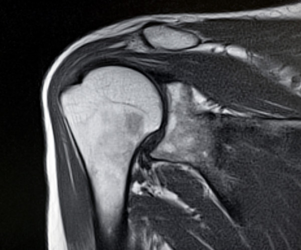
Suspected rotator cuff tear mri without contrast. The mri below also shows a tear in the rotator cuff as noted in the dark signal in the muscle white arrow. Ct arthrograms showing contrast white in the tear in the rotator cuff.
Some shoulder surgeons prefer mri shoulder arthrogram. The rotator cuff cannot be evaluated on a non contrast ct nor on a intravenous contrast ct. This type of test uses sound waves to produce images of structures within your body particularly soft tissues such as muscles and tendons.
When a full thickness tear is present in the rotator cuff mr arthrography will display the contrast solution extending through the defect in the cuff and into the subacromial subdeltoid bursa. If your doctor does recommend an mri for your shoulder there are two potential types of mri procedures that can be used to diagnose a rotator cuff tear.
Posterior Labrum Tear 3t Mri Arthrogram Shoulder Portland Mri
Rotator Cuff Injuries Often Diagnosed With Mri Diagnostic
 Example Of An Asymptomatic Patient With A Large Rotator Cuff Tear
Journal Of Lancaster General Health Depart3 V4i1
 Mr Arthrogram For Shoulder Microinstability And Hidden Lesions
 Rotator Cuff Injury Mri Practice Essentials Magnetic Resonance
 Mri Of The Shoulder Following Contrast Injection Directly Into The
 Rotator Cuff Tears Rotator Cuff 78 Steps Health Journal
 Rotator Cuff Injury Mri Practice Essentials Magnetic Resonance
 Rotator Cuff Mri Everything You Need To Know Dr Nabil
 Shoulder Mri Scan Cost Save 80 Off
 Full Text Partial Thickness Rotator Cuff Tears Clinical And
Https Encrypted Tbn0 Gstatic Com Images Q Tbn 3aand9gcrcb4nsg9pdslwdvkniqdzpgsw Zbredcyrrbpdnel8b7tzrp6v Usqp Cau
 Ultrasound Can It Replace Mri In The Evaluation Of The Rotator
 Shoulder And Elbow Surgery Shoulder Dislocation Resulting In
 Mri Rotator Cuff Injuries When You Need One When You Don T
 Role Of Conventional Mri And Mr Arthrography In Evaluating
 Rotator Cuff Tear Diagnosed By Conventional Direct Arthrography
 Role Of Conventional Mri And Mr Arthrography In Evaluating
 Pdf Efficacy Of Ultrasonography Guided Shoulder Mr Arthrography
 Mri Images Showing Torn Rotator Cuff
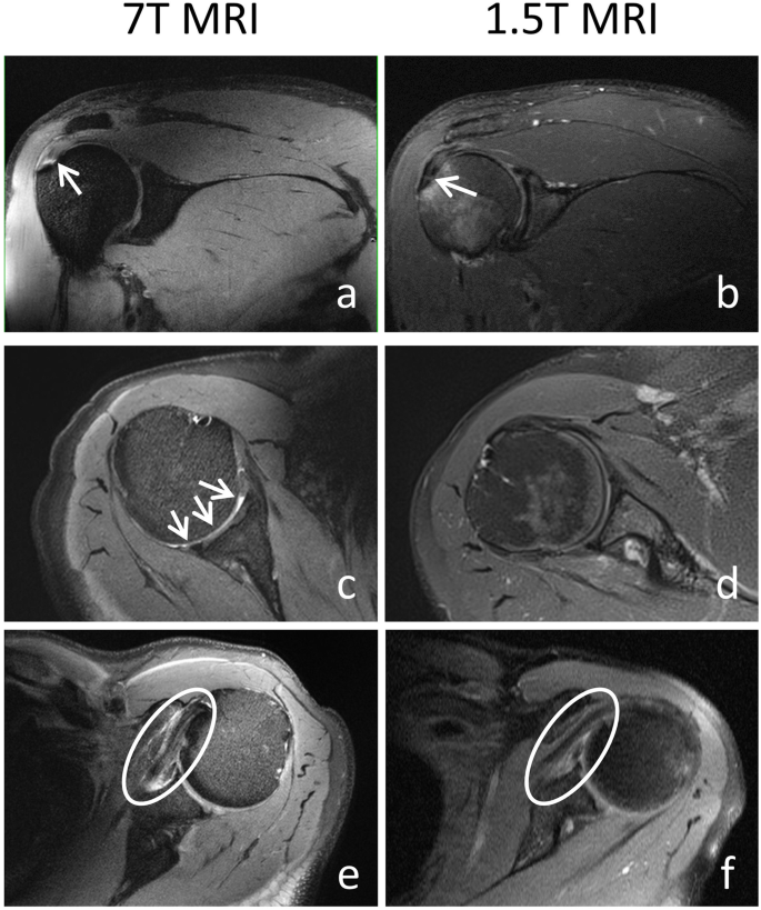 7 T Clinical Mri Of The Shoulder In Patients With Suspected
 Ultrasound Can It Replace Mri In The Evaluation Of The Rotator
Https Pubs Rsna Org Doi Pdf 10 1148 Rg 264055087
 How To Read Your Shoulder Mri Youtube
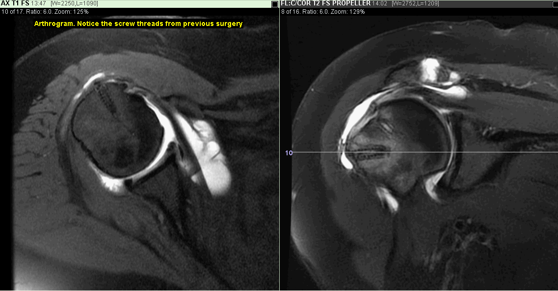 Mri Shoulder Scan Greater Waterbury Imaging Center
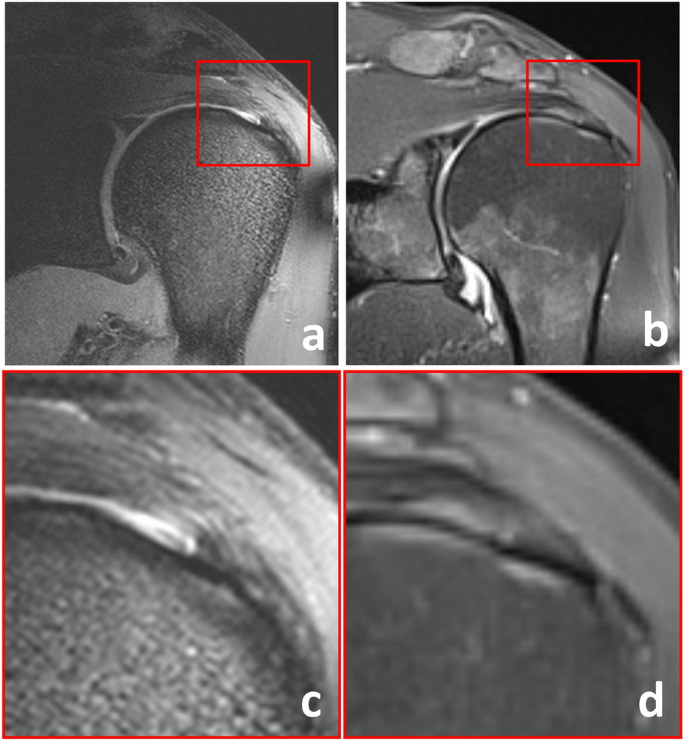 7 T Clinical Mri Of The Shoulder In Patients With Suspected
Posterior Labrum Tear 3t Mri Arthrogram Shoulder Portland Mri




Posting Komentar
Posting Komentar