Anterior Chamber Of Eye Anatomy
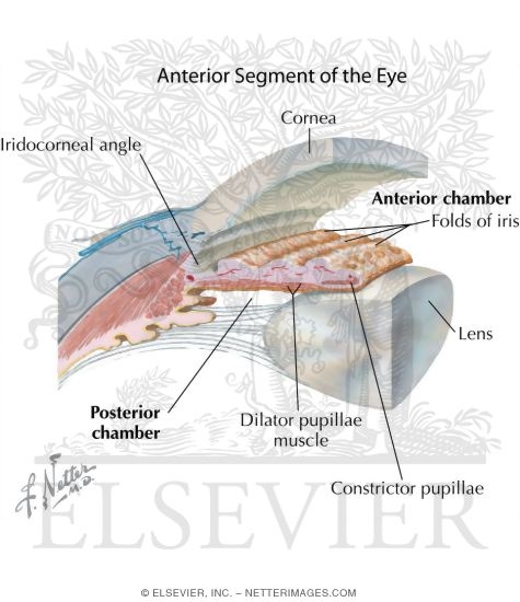
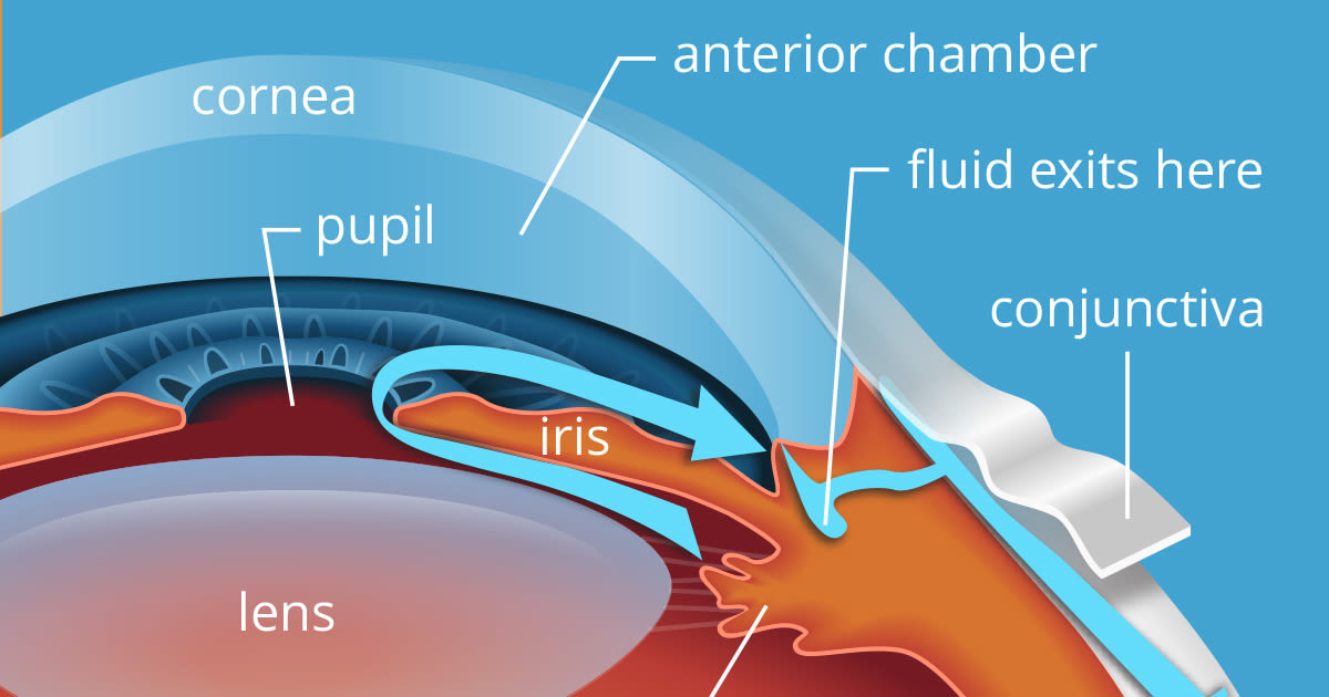
The fluid that fills this chamber is called the aqueous humor. The iris controls the amount of light that enters the eye by opening and closing the pupil.
 About Basic Eye Anatomy Gem Clinic Glaucoma Eye Management
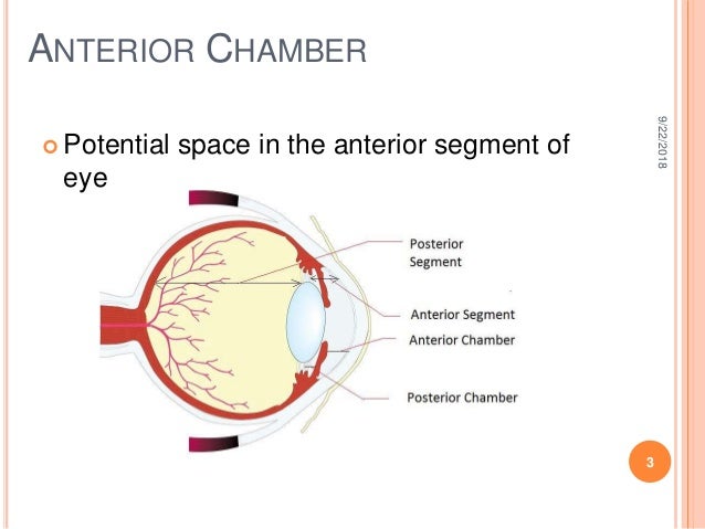
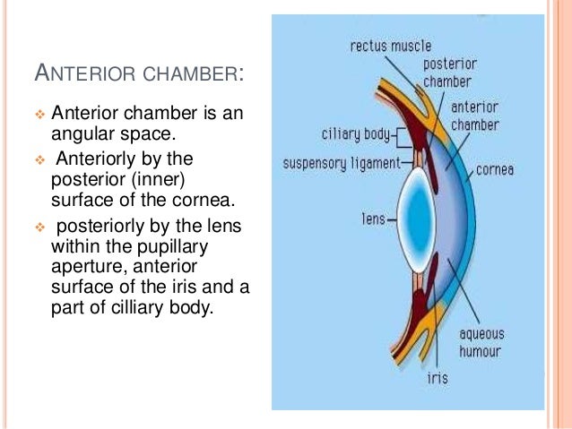
Posteriorly by the lens within the pupillary aperture anterior surface of the iris and a part of cilliary body.

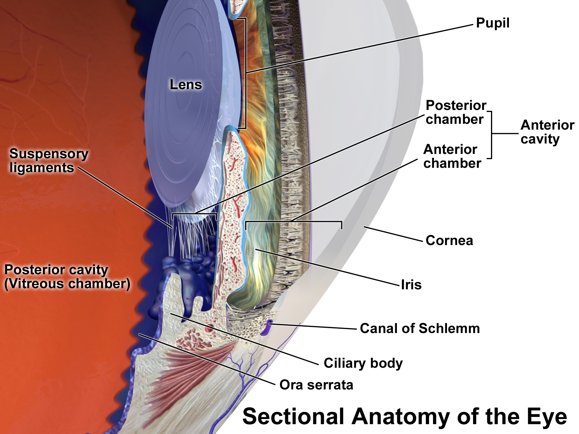
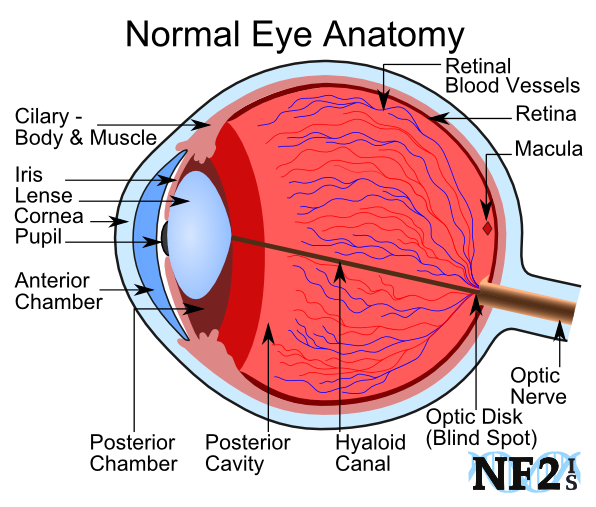
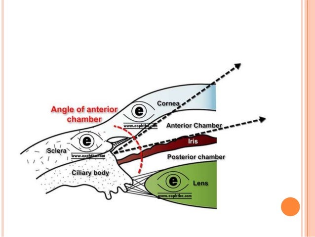
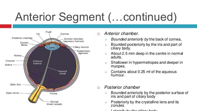
Anterior chamber of eye anatomy. The anterior chamber is the fluid filled space immediately behind the cornea and in front of the iris. The eye also contains three fluid filled chambers. Anterior chamber is an angular space.
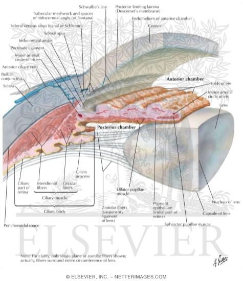
The aqueous humor helps to nourish the cornea and the lens. The volume between the cornea and the iris is known as the anterior chamber while the volume between the iris and the lens is know as the posterior chamber both chambers contain a fluid called aqueous humor. Anteriorly by the posterior inner surface of the cornea.
The inside of the eye is divided into three sections called chambers. The anterior chamber ac is the aqueous humor filled space inside the eye between the iris and the cornea s innermost surface the endothelium. In some eyes the ciliary body face is not visible being completely obscured by iris.
The degree to which the ciliary body face is visible depends on the level and angle of iris insertion. The iris uses muscles to change the size of the pupil. Aqueous humor is watery fluid produced by the ciliary body.
In hyphema blood fills the anterior chamber as a result of a hemorrhage most commonly after a blunt eye injury. The ciliary body face is the portion of the ciliary body that borders on the anterior chamber. Anterior chamber angle and the trabecular meshwork.
Hyphema anterior uveitis and glaucoma are three main pathologies in this area. We hope this picture anterior chamber and posterior chamber in the eye can help you study and research. For more anatomy content please follow us and visit our website.
We think this is the most useful. The anterior chamber is the front part of the eye between the cornea and the iris. The front section of the eye s interior where aqueous humor flows in and out providing nourishment to the eye.
A radial section of a portion of the retina reveals that the ganglion cells the output neurons of the retina lie innermost in the retina closest to the lens and front of the eye and the photosensors the rods and cones lie outermost in the retina against the pigment epithelium and choroid.
 Anterior And Posterior Chambers Of Eye Anatomy Of The Anterior Chamber
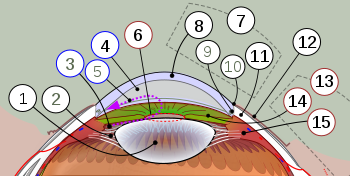 Anterior Segment Of Eyeball Wikipedia
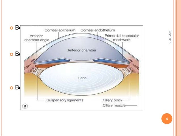 Anterior Chamber Anatomy Aqueous Production Drainage
 Major Ocular Structures Laramy K Independent Optical Lab
 Find Out How The Eye Works Fact Sheet Vision Eye Institute
Gross Anatomy Of The Eye By Helga Kolb Webvision
Eye Anatomy And How The Eye Works
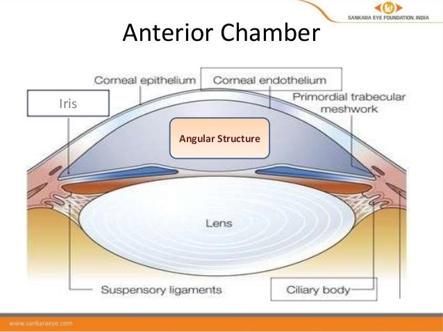 Anatomy Of The Anterior Chamber And Angle
 Anterior Chamber Anatomy Aqueous Production Drainage
 Anatomy Of Eye Flashcards Quizlet
 Aqueous Humour Physiology Britannica
 12 2 15 Va Ambulatory Report Pearls Anterior Chamber Uveitis
Stock Image Illustration Of The Eye Anatomy Closed Angle Cornea
 Anterior Chamber Of Eyeball Youtube
Ocular Anatomy Union Eye Works
The Anatomy Of The Eye Anterior Segment Precision Family Eye Care
 Anterior Chamber Of Eyeball Wikipedia
 Anatomy Of The Eye Columbia Eye Clinic
Duke Neurosciences Eye Ear Histology
 Anterior And Posterior Chambers Of Eye Anatomy Of The Anterior Chamber
 Anatomy Of The Eye And Trabecular Meshwork A Eye Diagram
 Anterior Chamber Of Eyeball Wikipedia
 Structure And Function Of The Eyes Eye Disorders Merck Manuals
 Eye Anatomy Amazing Point Of View Eye Care
 Anterior Chamber Of Eyeball Youtube
Eye Anatomy Ophthalmology Medbullets Step 2 3
Https Encrypted Tbn0 Gstatic Com Images Q Tbn 3aand9gcqcszjlt7dsxw6jag Hwh Bvvtaqmvshupepc00fsu4bvlzf7bk Usqp Cau
 Anterior Chamber And Posterior Chamber In The Eye
Anterior Segment Anatomy American Academy Of Ophthalmology
 Anterior Segment Anatomy American Academy Of Ophthalmology

Posting Komentar
Posting Komentar