Thoracic Spine X Ray Lateral
A thoracic spine x ray is an x ray of the 12 chest thoracic bones vertebrae of the spine. It is convex anteriorly the convexity of the lower three vertebrae being much greater than that of the upper two.
 Imaging The Cervical Thoracic And Lumbar Spine Radiology Key
How the test is performed the test is done in a hospital radiology department or in the health care provider s office.
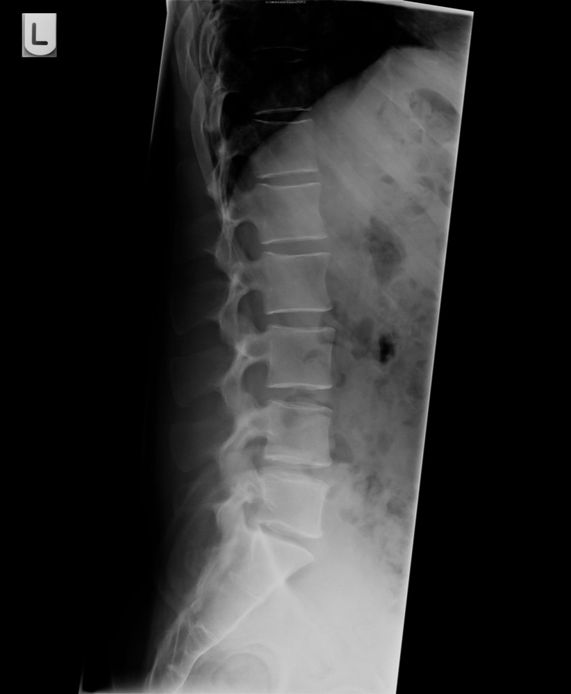
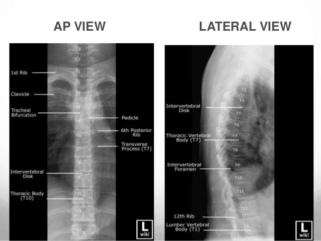
Thoracic spine x ray lateral. The thoracic spine ap view images the thoracic spine which consists of twelve vertebrae. A thoracic spine x ray is an imaging test used to inspect any problems with the bones in the middle of your back. Thoracolumbar spine x ray involves two views ap and lateral.
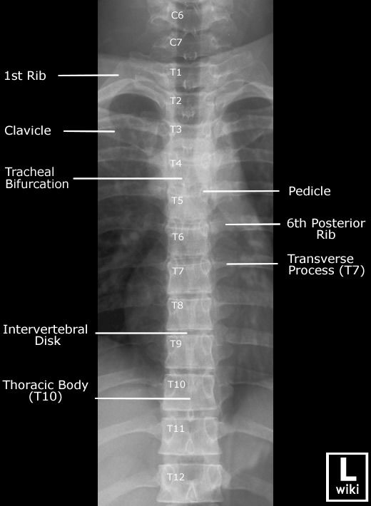
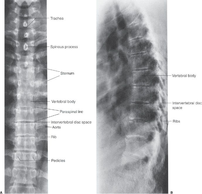
Images the entirety of the thoracic spine which consists of twelve vertebrae. In addition to the thoracic vertebral bodies spinous processes and transverse processes a good quality thoracic spine ap projection shows the left ventricle gastric bubble right left hemidiaphragm posterior ribs and right left clavicle. Intervertebral joints are seen in profile.
It is utilized in many imaging contexts including trauma postoperatively and for chronic conditions. Check it s an adequate view for a lumbar spine view you should be able to see l1 l5 but also the full t12 vertebral body t11 12 and the sacrum on the ap view. The lumbar spine is made up of five vertebral bones.
A lumbosacral spine x ray or lumbar spine x ray is an imaging test that helps your doctor view the anatomy of your lower back. Lateral lumbar x ray of a 34 year old male the lumbar curve is more marked in the female than in the male. Intervertebral joints and neural foramen are open with the superimposition of the posterior spinous processes.
Structure is function in the next few weeks we will be adding a low radiation digital x ray system to our office. An x ray uses small amounts of radiation to see the organs tissues and bones of. It begins at the middle of the last thoracic vertebra and ends at the sacrovertebral angle.
Patient position the patient is erect or supine d. The lateral projection requires the upper limbs to be removed from the path of the direct x ray beam minimizing the superimposition of the proximal humeri over the thoracic vertebrae in all variations of positioning the humeri are extended 90º to the thorax with the elbows flexed so that the forearms are parallel to the thorax. Often performed erect unless otherwise indicated.
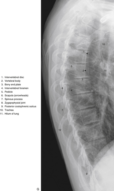
The vertebrae are separated by flat pads of cartilage called disks that provide a cushion between the bones. Many of you have been with our practice for some time and remember the x ray analysis we had as a part of our new patient exam. The lateral projection shows the left and right hemidiaphragm vertebral bodies intervertebral disc spaces and posterior ribs.
The sacrum is.

 Thoracic Spine Radiology Tutorial Youtube

 Thoracic Spine Flashcards Quizlet
Https Encrypted Tbn0 Gstatic Com Images Q Tbn 3aand9gcr3nfouchp5gkq7f6rmqjucscokfzaonvriccugzpucsw 1wz1m Usqp Cau
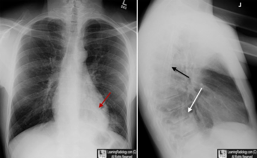
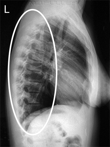
 Lateral Chest Xrays Of Patients Showing The Normal Thoracic Spine
 X Ray Positioning For Thoracic Spine Youtube
 Ce4rt Radiographic Positioning Of The Thoracic Spine For X Ray Techs
 Thoracic Spine Compression Fractures Vertebra Plana
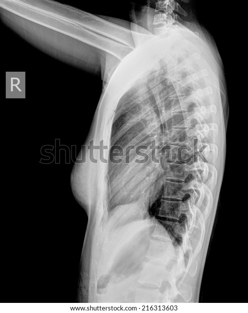 Film Xray Women Upper Thoracic Spine Stock Photo Edit Now 216313603
 Thoracic Spine Lateral Canine Veterinary X Ray Positioning Guide

 Normal Thoracic Spine Radiology Case Radiopaedia Org
A And B Posteroanterior And Lateral X Ray Image Of The Thoracic
 Thoracic Spine Ap View Radiology Reference Article
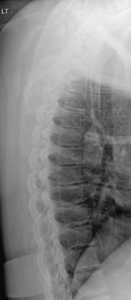
 Lateral Thoracic Spine Xray Radiology X Ray Radiology Student
 Diffuse Idiopathic Skeletal Hyperostosis Wikipedia
 A Ap And Lateral Views Of The Thoracic Spine Demonstrated
 Initial Plain X Rays Of The Thoracic Spine Showing That The
 Normal Lateral Thoracic Spine Radiograph Radiology Case
The Thoracolumbar Spine
 Radiology For Anatomists Thoracic Spine Oblique

 Normal Lateral Thoracic Spine Radiograph Radiology Case
 Anteroposterior And Lateral X Rays Of The Thoracic Spine

 The Thoracic Region Basicmedical Key
 Lateral X Ray Of The Thoraco Lumber Spine Showing Compression
 9 Spine And Pelvis Radiology Key
 Lateral X Ray Thoracic Spinal Instrumentation Stock Image C043
 Normal Thoracic Spine X Ray 9 Year Old Radiology Case

 Getting A Good View Of The Cervicothoracic Junction Swimmer S
 Thoracic Spine Radiology Key
 Radiography Of Chest And Spine
 The Thoracolumbar Spine
Posting Komentar
Posting Komentar