Rotator Cuff Tear Mri Arthrogram
In diagnosing a full thickness tear or a partial thickness rotator cuff tear mr arthrography is more sensitive and specific than either mri or ultrasound p 0 05. With this kind of test a contrasting dye is injected into the shoulder though this dye is different than that used in a standard arthrogram.
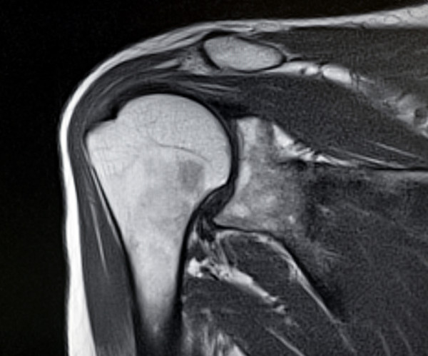
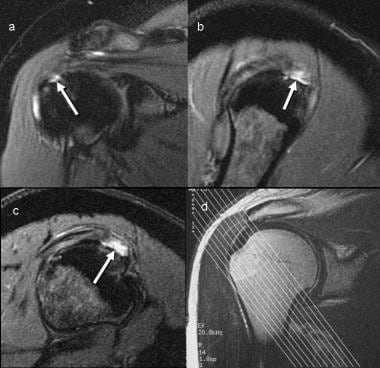
 A The Oblique Coronal T1 Weighted Image Of Magnetic Resonance
The superior aspect of this outlet the coracoacromial arch includes the acromion which is an anterolateral extension of the scapular spine the anterior third of the.

Rotator cuff tear mri arthrogram. Tendon retraction may also be present which can be graded using the patte classification. The rotator cuff tendons are fairly thick tendons about as thick as your little finger and wide each one about as wide as three of your fingers. A routine shoulder mri which takes about 25 minutes of actual scan time and shoulder mr arthrogram which involves an x ray procedure during which contrast is injected directly into the.
Rotator cuff these are a set of muscles which start on the shoulder blade but attach to the arm bone the humerus by tendons see patient guide to rotator cuff tendinitis. Then the mri is performed. Although a rotator cuff tear won t show up on an x ray this test can visualize bone spurs or other potential causes for your pain such as arthritis.
This type of test uses sound waves to produce images of structures within your body particularly soft tissues such as muscles and tendons. The rotator cuff cannot be evaluated on a non contrast ct nor on a intravenous contrast ct. To analyze a rotator cuff tear an mri would usually performed unless it is contraindicated such as by a pacemaker.
There are no significant differences in either sensitivity or specificity between mri and ultrasound in the diagnosis of partial or full thickness rotator cuff tears p 0 05. A ct arthrogram should be specified in order to rule out a rotator cuff tear. Diagnosing a rotator cuff tear or other type of tissue tear isn t always as simple as a patient may think even if the resulting shoulder pain is classical of this condition.
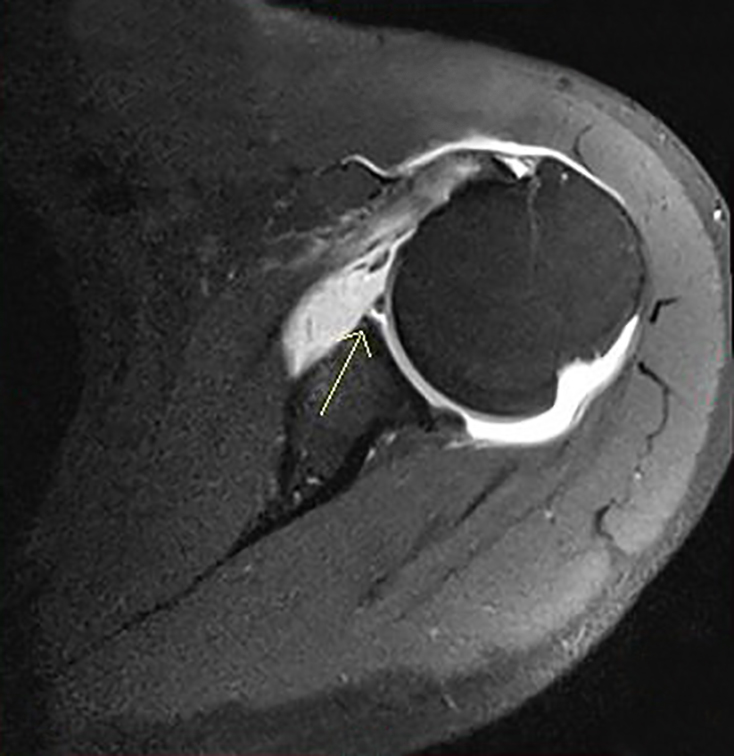
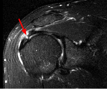
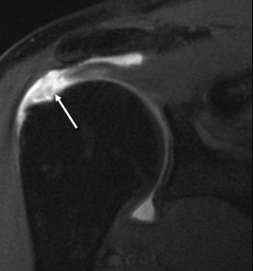
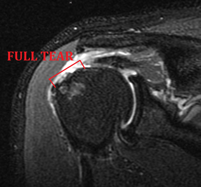
The presence of a tendon defect filled with fluid is the most direct sign of rotator cuff tear. In some cases an arthrogram can be done using the same technology as a mri radiologyinfo explains. The subacromial bursa serves as a buffer between the rotator cuff and coracoacromial arch and is located below the acromion coracoacromial ligament and above the rotator cuff.
The contrast dye helps outline the muscles and tendons within the rotator cuff. If your doctor does recommend an mri for your shoulder there are two potential types of mri procedures that can be used to diagnose a rotator cuff tear. Indirect signs on mri are subdeltoid bursal effusion medial dislocation of biceps fluid along biceps tendon and diffuse loss of peribursal fat planes.
Find out which procedure the mr arthrography or a standard mri is better at detecting a rotator cuff tear.
 Mri Arthrogram Glenoid Labral Tear Medical Imaging Mri Radiology
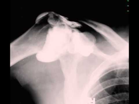 Rotator Cuff Tear On Shoulder Arthrogram Youtube
 Cedars Sinai
 Mr Arthrogram For Shoulder Microinstability And Hidden Lesions
 Cedars Sinai

 Aim Offers Mri Arthrograms Aim Medical Imaging

 Rotator Cuff Tears Shoulder Elbow Orthobullets
Https Www Ajronline Org Doi Pdfplus 10 2214 Ajr 08 1097
 Cedars Sinai
Rotator Cuff Surgery Sports Medicine Seattle Wa
Role Of Conventional Mri And Mr Arthrography In Evaluating
 Rotator Cuff Tear Of The Shoulder Ct Arthrogram Finding Youtube
 Rotator Cuff Tear Diagnosed By Conventional Direct Arthrography
 Ge Healthcare
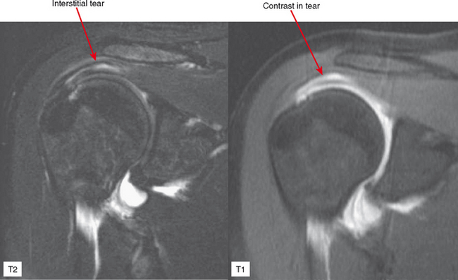
 Rotator Cuff Pitfalls Radsource
 Cedars Sinai
 Normal Shoulder Arthrogram Radiology Case Radiopaedia Org

 Cedars Sinai
 Full Text Partial Thickness Rotator Cuff Tears Clinical And
 Mr Arthrogram For Joints Shoulder Elbow Wrist Hip Knee Ankle
 Shoulder Mri


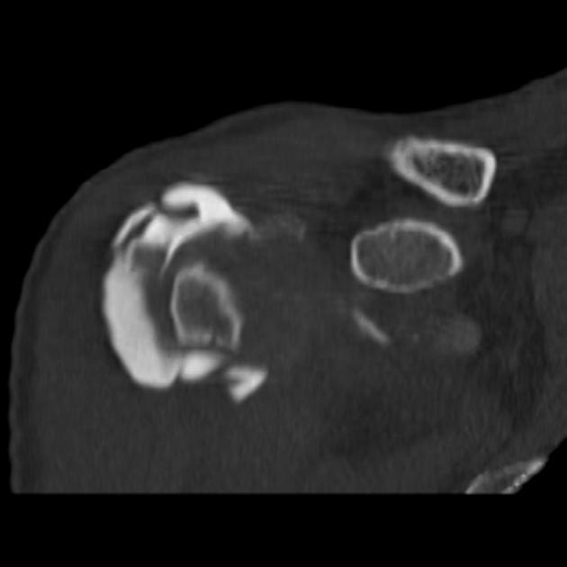 Rotator Cuff Tear Ct Arthrography Radiology Case Radiopaedia Org
 Rotator Cuff Tear And Impingement Syndrome Ct Arthrogram Finding
 Arthrography And Joint Injection And Aspiration Principles And
Https Encrypted Tbn0 Gstatic Com Images Q Tbn 3aand9gcrcb4nsg9pdslwdvkniqdzpgsw Zbredcyrrbpdnel8b7tzrp6v Usqp Cau
 Figure 3 From Management Of The Throwing Shoulder Cuff Labrum
 Rotator Cuff Pitfalls Radsource
 Rotator Cuff Tears Shoulder Elbow Orthobullets

 Rotator Cuff Injury Mri Practice Essentials Magnetic Resonance
 Rotator Cuff Injury Mri Practice Essentials Magnetic Resonance
 Cedars Sinai
 Rotator Cuff Tear Radiology Reference Article Radiopaedia Org
 Atlanta Rotator Cuff Tear Shoulder Surgery L Bone Joint Doctor
Posting Komentar
Posting Komentar