What Color Is Cancer On An Ultrasound
2010 epub ahead of print. What color does cancer show up on ultrasound.

Often during surgery to remove the tumor from your colon.
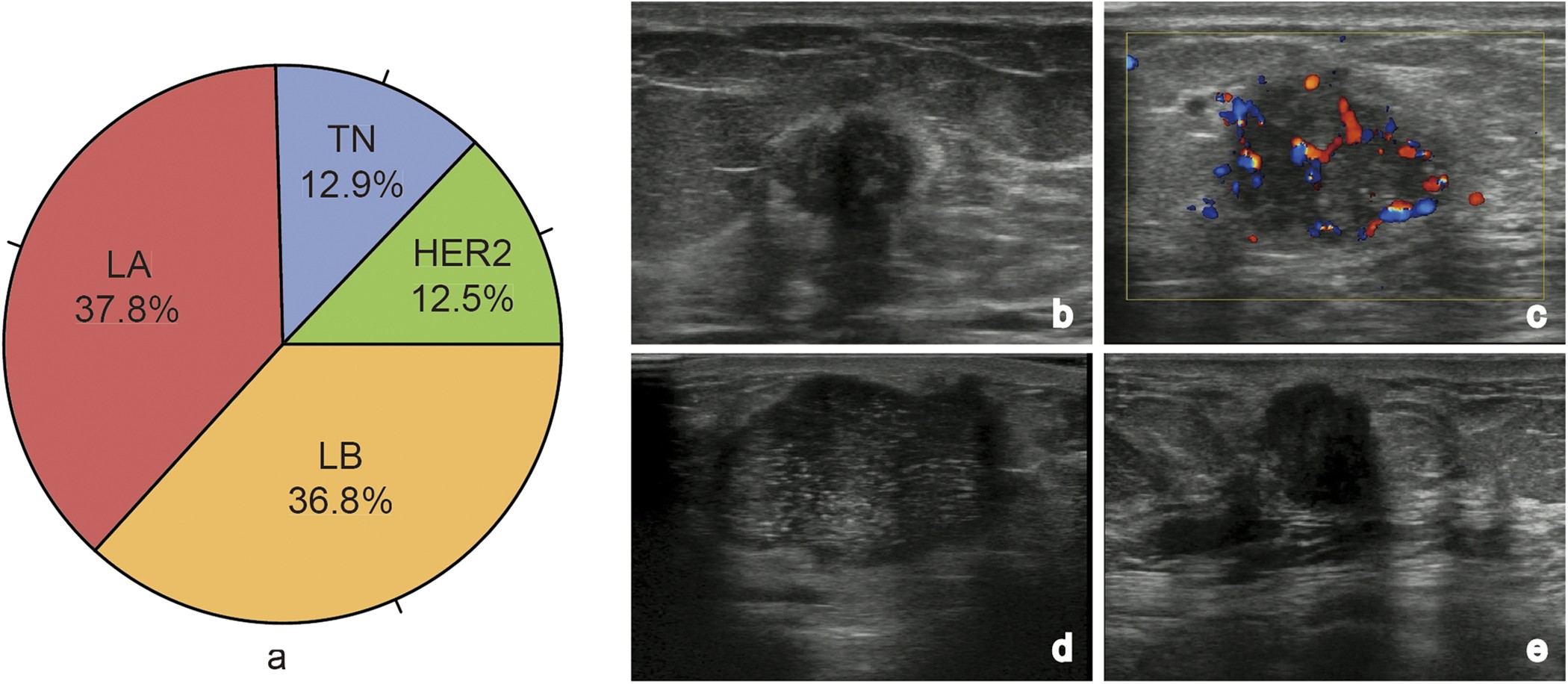
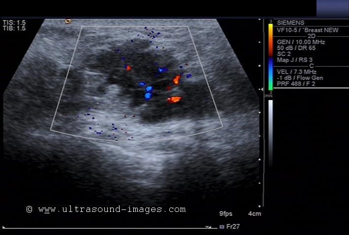
What color is cancer on an ultrasound. The use of breast ultrasound color doppler vascular pattern morphology improves diagnostic sensitivity with minimal change in specificity. Schulman h conway c zalud i et al. It is important to remember that not all densities or microcalcifications seen on mammography are cancer.
A 14 year old female asked. Color doppler has made it easier for doctors to find out if cancer has spread into blood vessels especially in the liver and pancreas. An example of early signs that may not show up on ultrasound are tiny calcium deposits called microcalcifications.
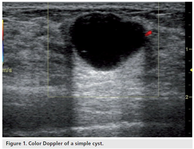
Cysts tumors and growths will appear dark on the scan. These images can show the. It can show whether the cancer has spread inside your pelvis or to your liver.
An ultrasound machine has 3 key parts. Prevalence in a volunteer population of pelvic cancer detected with transvaginal ultrasound and color flow doppler. 4d ultrasound can gather 3d volumes and represents the difference between video and a still.
This test uses sound waves to make a picture of your organs. Have particularly dense breast tissue. Bildgebung 61 4 291 294 1994.
Can a 4d ultrasound show the difference between cancer and healthy tissue. Sometimes cancers show both features. Just because there is a dark spot on your ultrasound doesn t mean that you have breast cancer.
How does it work. This is because it may miss some early signs of cancer. On a mammogram cancer can look like a white irregular density or can appear as small specks of calcium microcalcifications clumped in a particular pattern.
Sohn c baudendistel a bastert g. Ultrasound may be used if you. Diagnosis of the breast tumor entity with vocal fremitus in ultrasound diagnosis.
32 years experience in radiation oncology. Breast ultrasound is not usually done to screen for breast cancer. A control panel a display screen and a transducer which usually looks a lot like a microphone or a computer mouse.
The images from a breast ultrasound are in black and white.
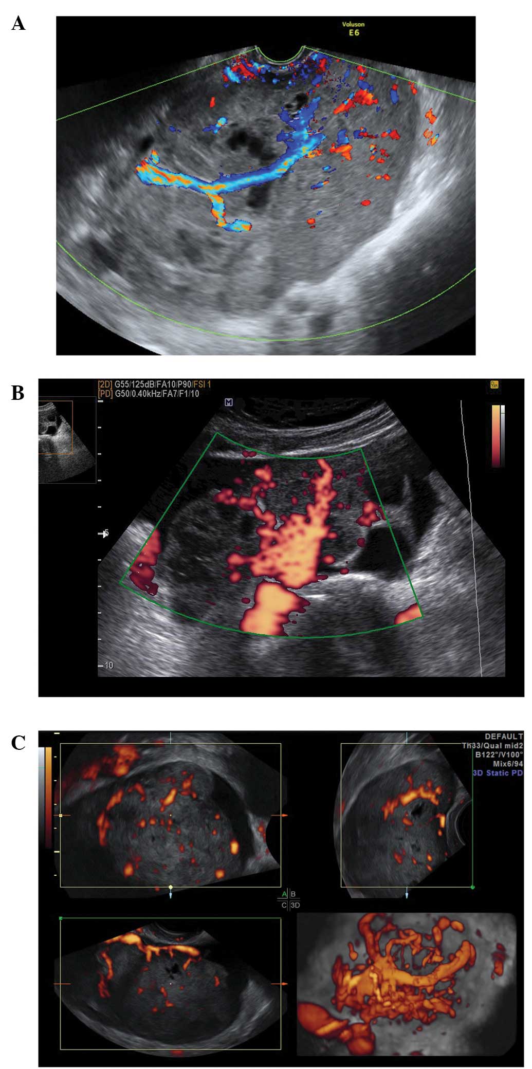
 Ultrasound Characteristics Of Endometrial Cancer As Defined By International Endometrial Tumor Analysis Ieta Consensus Nomenclature Prospective Multicenter Study Epstein 2018 Ultrasound In Obstetrics Amp Gynecology Wiley Online Library
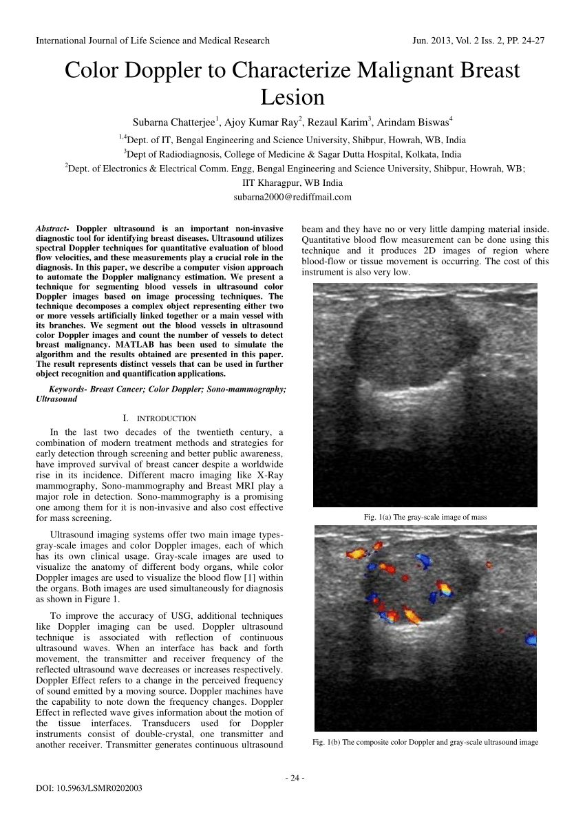 Pdf Color Doppler To Characterize Malignant Breast Lesion
 Staging Of Breast Cancer With Ultrasound Sciencedirect
 Ultrasound Doppler Principles Preparation Results And More

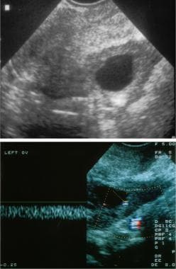
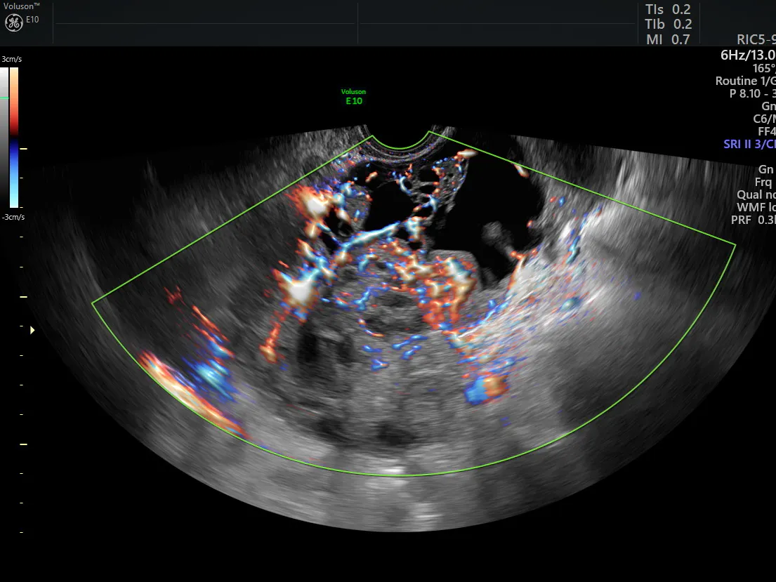
![]() Transvaginal Color Doppler Ultrasound Showing A Cervical Cancer With Download Scientific Diagram
Transvaginal Color Doppler Ultrasound Showing A Cervical Cancer With Download Scientific Diagram
![]() Transvaginal Color Doppler From A Cervical Cancer Showing An Abundant Download Scientific Diagram
Transvaginal Color Doppler From A Cervical Cancer Showing An Abundant Download Scientific Diagram
 Can An Ultrasound Detect Cancer Moffitt
Https Encrypted Tbn0 Gstatic Com Images Q Tbn 3aand9gcsal2b6q9sttsrad3nul Zk8z1gz03evajv7lgvikjsmq4dyfs4 Usqp Cau
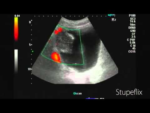 Color Doppler Ultrasound Bladder Cancer Youtube
 A Gallery Of High Resolution Ultrasound Color Doppler 3d Images Breast
 Diagnostic Value Of Ultrasound In Metastatic Liver Cancer Abdominal Key
 Ultrasound And Color Doppler Ultrasound Of Soft Tissue Tumors And Tumorlike Lesions Radiology Key
 Gray Scale And Color Doppler Ultrasound Characteristics Of Endometrial Cancer In Relation To Stage Grade And Tumor Size Epstein 2011 Ultrasound In Obstetrics Amp Gynecology Wiley Online Library
 Ultrasound Imaging Cancerquest
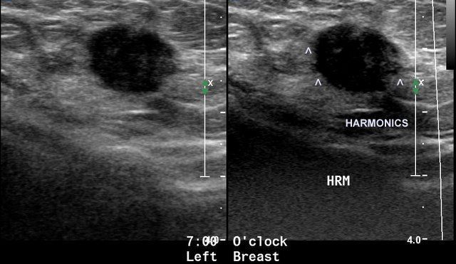
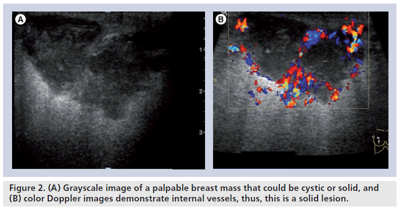 Color Doppler Sonography Characterizing Breast Lesions
 Color Doppler Sonography Characterizing Breast Lesions
![]() Eposters Use Of Transabdominal Color Doppler Ultrasound For Detection Of Colon Cancer In Patients With Nonspecific Abdominal Symptoms A Personal Experience And Meta Analysis Of The Literature
Eposters Use Of Transabdominal Color Doppler Ultrasound For Detection Of Colon Cancer In Patients With Nonspecific Abdominal Symptoms A Personal Experience And Meta Analysis Of The Literature
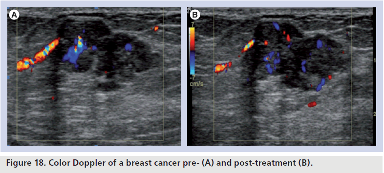
 Figure 1 From Th Does Color Doppler Ultrasound Vascularity Predict The Response To Neoadjuvant Chemotherapy In Breast Cancer Semantic Scholar
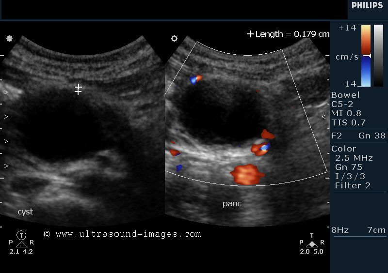 A Gallery Of High Resolution Ultrasound Color Doppler 3d Images Pancreas
 Color Doppler Sonography Characterizing Breast Lesions
Malignant Ovarian Tumor Imaging Practice Essentials Computed Tomography Magnetic Resonance Imaging
 Color Doppler Sonography Characterizing Breast Lesions
 Color Doppler Sonography Characterizing Breast Lesions
 The Role Of Breast Ultrasound In Early Cancer Detection Sciencedirect
 Neoangiogenesis In Early Cervical Cancer Correlation Between Color Doppler Findings And Risk Factors A Prospective Observational Study World Journal Of Surgical Oncology Full Text
 Table 1 From Th Does Color Doppler Ultrasound Vascularity Predict The Response To Neoadjuvant Chemotherapy In Breast Cancer Semantic Scholar
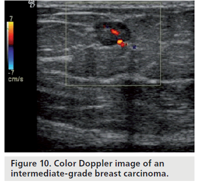
 5 2 4 Breast Carcinoma Diagnostic Criteria Color Doppler Ultrasound Cases
 Sonocine Sonogram Results For Breast Cancer Detection Youtube
 Ultrasound And Color Doppler Imaging Ultrasound Mens Tops Mens Tshirts
 Selection Of Hens With Normal Ovaries Or Ovarian Tumors Using A C Download Scientific Diagram
 The Characteristic Ultrasound Features Of Specific Types Of Ovarian Pathology Review
 Color Doppler Sonography Characterizing Breast Lesions
:max_bytes(150000):strip_icc()/breast-cancer-tumors-what-are-they-430277-v12-d91aad27f20b4f06aae6afc5a55868da.png) Breast Masses Cancerous Tumor Or Benign Lump
Breast Masses Cancerous Tumor Or Benign Lump
 Identifying Ultrasound And Clinical Features Of Breast Cancer Molecular Subtypes By Ensemble Decision Scientific Reports
 Imaging The Suspected Ovarian Malignancy 14 Cases Mdedge Obgyn
 Diagnostic Value Of Ultrasound In Metastatic Liver Cancer Abdominal Key
 A Gallery Of High Resolution Ultrasound Color Doppler 3d Images Breast
Posting Komentar
Posting Komentar