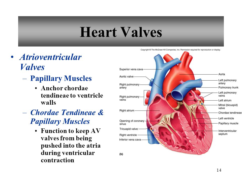
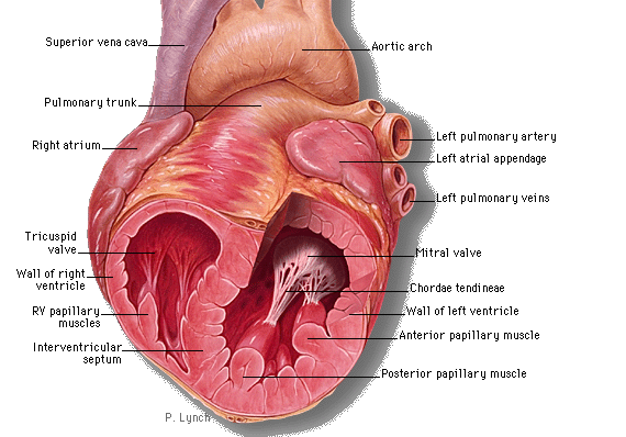
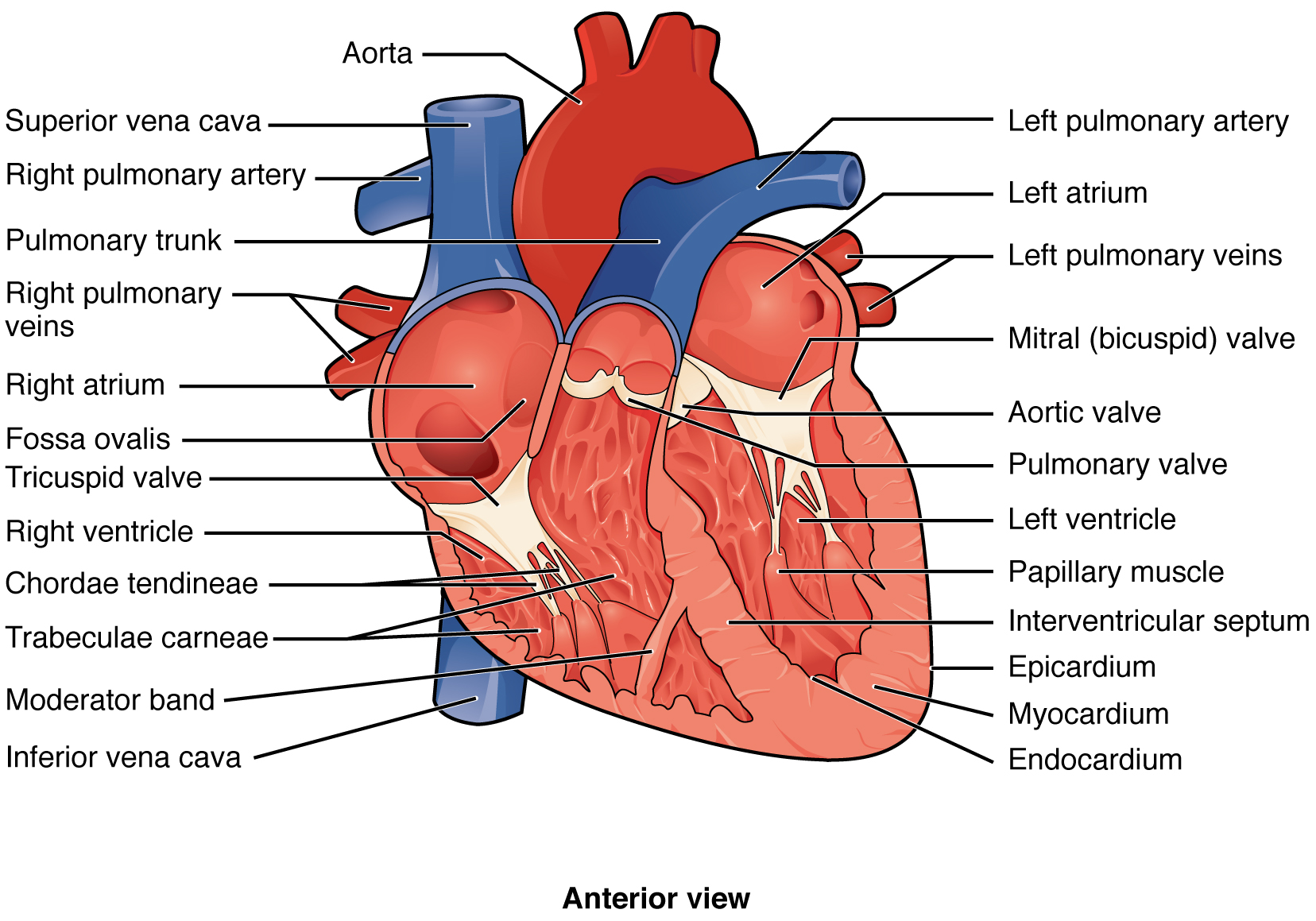
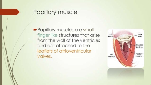
Function Of Papillary Muscles
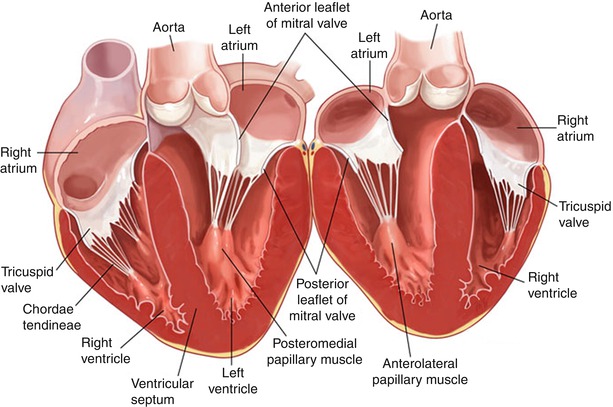
The remaining three septal posterior and anterior muscles can be found in the walls of the right ventricle and connect to the. The papillary muscles pull the chordae tendineae taut during ventricular contraction which prevents prolapse of the atrioventricular valves into the atria.
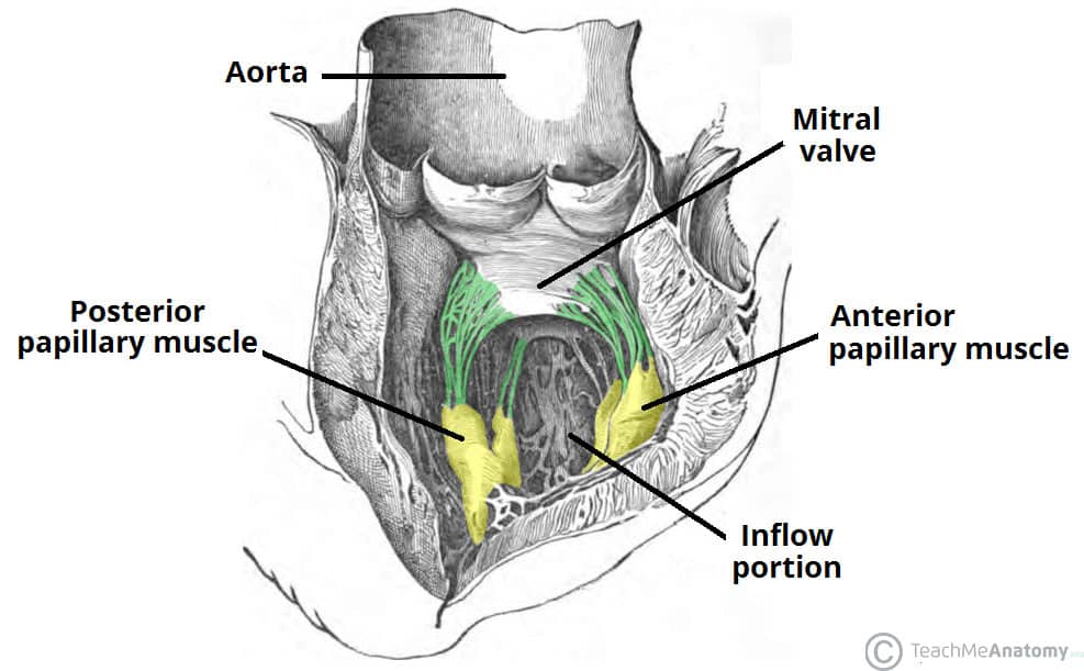 The Heart Valves Tricuspid Aortic Mitral Pulmonary Teachmeanatomy
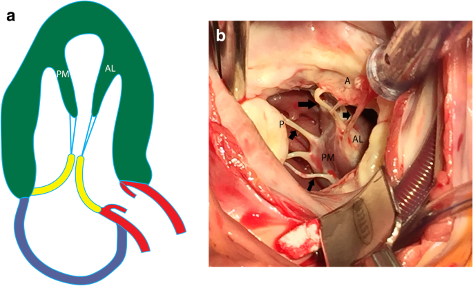
Prevents prolapse of anterior and posterior cusps of mitral valve during systole.
Function of papillary muscles. Importance in cardiovascular diseases. A small muscle within the heart that anchors the heart valves. The papillary muscles constitute about 10 of the total heart mass.
The role of the papillary muscle is to prevent inversion or prolapse or the atrioventricular valves. The papillary muscles are vital structures within the heart that are located in the left and right ventricles the two lower heart chambers. Function of the papillary muscles cannot be seriously impaired or blood flow through the heart is.
How many papillary muscles are there. The papillary muscles are muscles located in the ventricles of the heart. The anchor ropes are the chordae tendineae thread like bands of fibrous tissue that attach on one end to the edges of the tricuspid and mitral valves of the heart and on the other end to the papillary muscles.
The papillary muscles are muscles located in the ventricles of the heart. Dysfunction of a papillary muscle would prevent some of the chordae from functioning properly which could lead to valvular prolapse. The muscles of the right ventricles three in number arise in the ventricular wall.
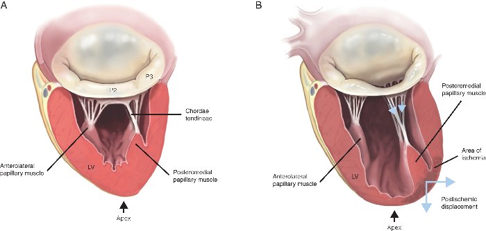
Mitral valve insufficiency due to dysfunction or rupture of papillary muscle coronary artery disease with papillary muscle ischemia and or infarction. The muscles anchor themselves to the cusps or flaps of the atrioventricular valves such as the tricuspid and mitral valve through the chordae tendineae. There are five papillary muscles which attach to the heart s atrioventricular valves.
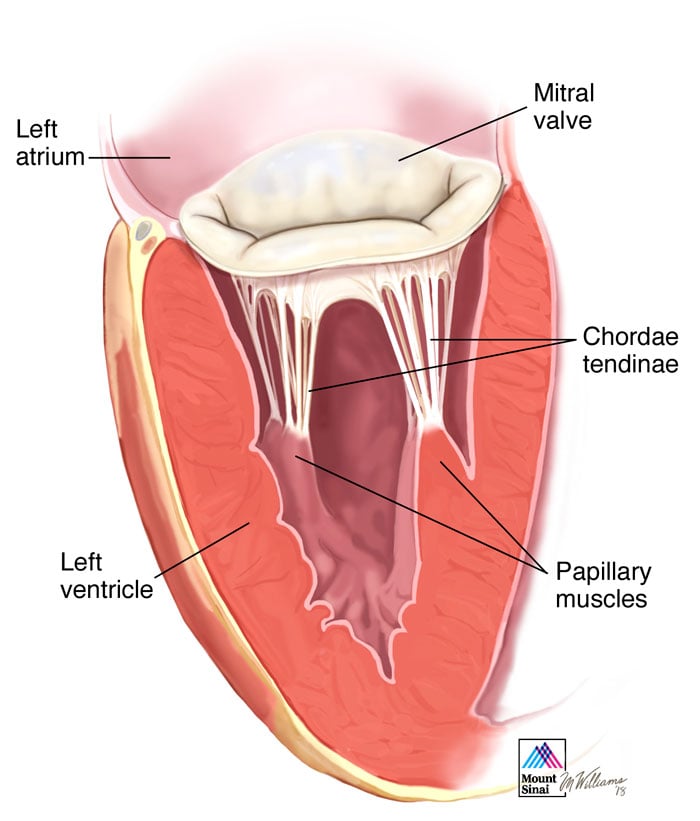
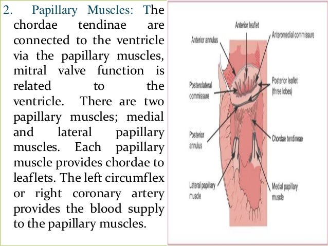
They perform important and necessary work allowing blood to flow from the atria to the ventricles. Subvalvular apparatus of the mitral valve chordae tendinae and papillary muscles because the annulus resides in the left atrio ventricular groove and the chordae tendinae are connected to the ventricle via the papillary muscles mitral valve function is integrally related to the ventricle. They attach to the cusps of the atrioventricular valves also known as the mitral and tricuspid valves via the chordae tendineae and contract to prevent inversion or prolapse of these valves on systole or ventricular contraction.
Two of the muscles are located in the posterior and anterior walls of the left ventricle and connect to the two cusps of the bicuspid valve. As aforesaid these muscles can be found in the heart.
 Chordae Tendineae Wikipedia
 Characterization Of 3 Dimensional Papillary Muscle Displacement In In Vivo Ovine Models Of Ischemic Functional Mitral Regurgitation Sciencedirect
 Functional Mr Structural Heart Research Innovation Laboratory
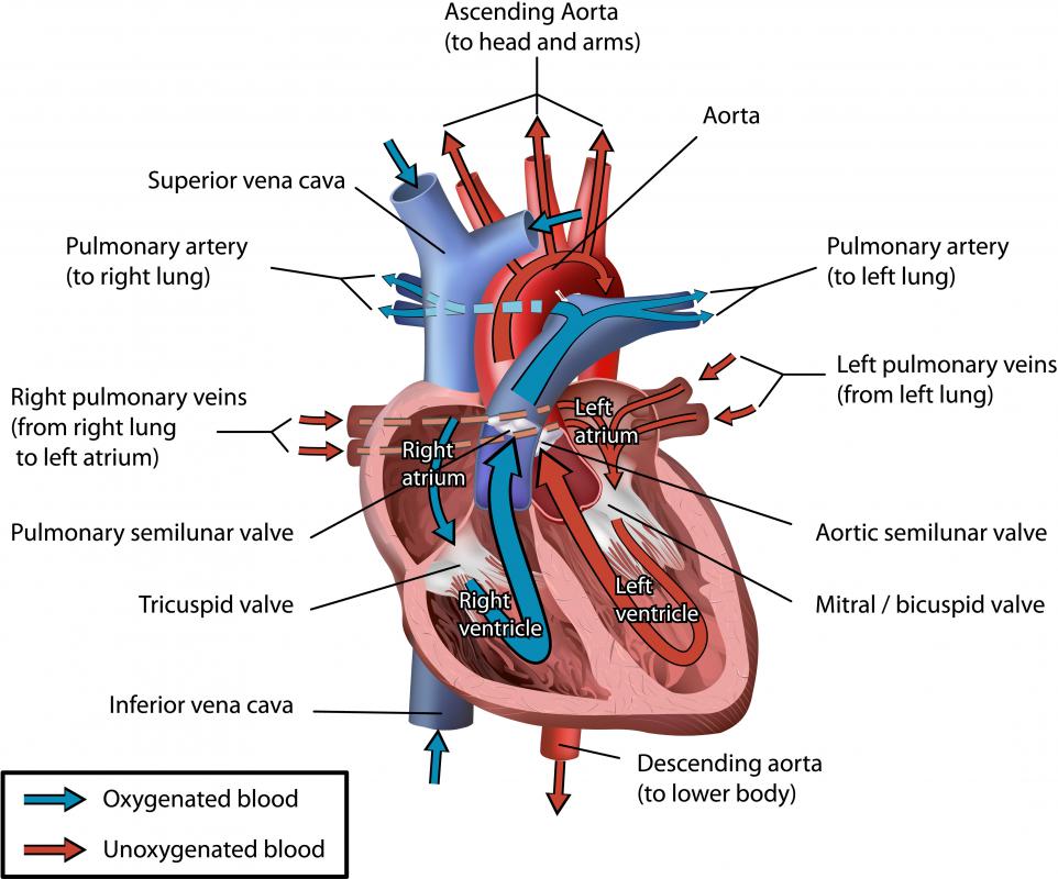
 The Heart As A Pump And Function Of The Heart Valves
 Mitral Valve Anatomy Name 5 Components Mitral Valve Cardiac Sonography Echocardiogram
Https Encrypted Tbn0 Gstatic Com Images Q Tbn 3aand9gcq Oeczm5n4bu7lb5mkphmro6qainedg1a0 Ny Aot0fmezjhx Usqp Cau
Https Uknowledge Uky Edu Cgi Viewcontent Cgi Article 1137 Context Me Etds
Papillary Muscles And The Left Ventricle Mitral Valve Repair Center
 What Are The Papillary Muscles With Pictures
 What Are The Papillary Muscles Mvp Resource
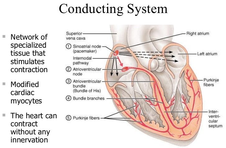 Purkinje Fibers Anatomy Location Function Anatomy Info
 Papillary Muscles Are Found In Mammalian Toppr Com
 What Is Function Of Papillary Muscle Cardioonline Basic And Advanced Cardiovascular Medicine Cariology Concepts And Review Dr Nabil Paktin Md Facc دکتـور نبــــیل پاکطــــین
 Cardiovascular System Ppt
 Ch18 Valves Of The Heart

 1 Chapter 15 Cardiovascular System 2 Size Of Heart Average Size Of Heart 14 Cm Long 9 Cm Wide Grams About Size Of Your Closed Fist Ppt Download

 Anatomy Block 1 Heart Flashcards Quizlet
 Magnetic Resonance Imaging Of The Papillary Muscles Of The Left Ventricle Normal Anatomy Variants And Abnormalities Springerlink
 Trabeculae Carneae Wikipedia
 Papillary Rupture
 Anatomy Block 1 Heart Flashcards Quizlet
 A Contractile Function In Papillary Muscle Fibers From 5 And Download Scientific Diagram
 Chordae Tendineae Heart
 Papillary Muscle Wikipedia
 What Are The Papillary Muscles Mvp Resource
 Anatomy Of The Tricuspid Valve And Pathophysiology Of Functional Tricuspid Regurgitation Thoracic Key
 Pin On Health
 Anatomy Of The Tricuspid Valve
 Interior Of Heart Chorae Tendineae Papillary Muscles Trabeculae Carneae Semilunar Valve B Heart Diagram Cardiovascular System Human Anatomy And Physiology
 What Is The Mitral Apparatus Mvp Resource
 Papillary Muscle Wikipedia
 Principles Of Carpentier S Reconstructive Mitral Valve Surgery Thoracic Key
 Papillary Muscles Of Left Ventricle Morphological Variations And It S Clinical Relevance Sciencedirect
Posting Komentar
Posting Komentar